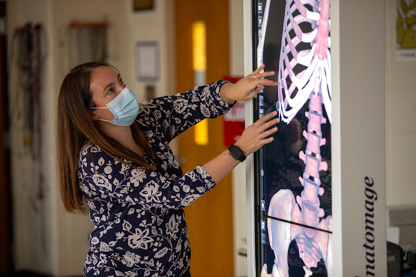
“I believe it will keep students engaged and interested, which can help them retain what they learn in the lab.”
The whole world has gone virtual. However, in the science community, technology has always been forging ahead, not out of pandemic necessity, but as a tool to advance learning.
At Immaculata, students in anatomy and physiology classes now have the benefit of learning through virtual dissection of human cadavers. In January of 2020, the university secured a grant from the Henry A. Quinn Charitable Foundation to purchase an Anatomage Table, which is the world’s first virtual dissection table.
Featuring the most advanced 3-D anatomy visualization system, students can see anatomy on the table exactly as they would on a cadaver. According to Kelly Orlando, Ph.D., associate professor of biology, the table also contains MRI and CT scans, as well as access to clinical cases to expose students to anatomical variations and diseased states. “They will be able to visualize physiological processes such as the pumping of the heart, and I can set up lab practical-type activities on the table so that students can explore the organ systems that we are studying,” Orlando explains. “I believe it will keep students engaged and interested, which can help them retain what they learn in lab.”
The Anatomage Table is a critical element for the new Anatomy and Physiology Lab, which will be a central component of the Parsons Science Pavilion. The table will augment curriculum for students in a number of programs including nursing, nutrition, biology, exercise science, allied health and even music therapy. The table’s digital library also contains a number of animal scans, which will be of use for pre-veterinary students.